
_1_edited.png)
INTRA-ABDOMINAL INFECTIONS
Intra-abdominal infections (IAIs) describe and is a broad term that encompasses a diverse number of infectious and disease processes. IAIs represent one of the most frequent gastrointestinal emergencies and a serious cause of morbidity and mortality.
IAIs are broadly defined as peritoneal inflammation in response to microorganisms, resulting in purulence (containing or forming pus) in the peritoneal cavity. A traditional classification universally accepted, divides intra-abdominal infections into complicated and uncomplicated based on the extent of infection.
Uncomplicated abdominal infections involve inflammation within the walls of the gastrointestinal (GI) tract and only involve a single organ and do not extend to the peritoneum. They are often simple to treat; however, when treatment is delayed or inappropriate, or the infection involves a more virulent nosocomial microbe, the risk of progression into a complicated abdominal infection, significantly increases.
Complicated abdominal infections extend beyond the source organ into the peritoneal space. They cause peritoneal inflammation and are associated with localized or diffuse peritonitis.
CONTENTS
-
Bile ducts and the human liver
-
Overview
-
Symptoms of cholangitis
-
Treating cholangitis
-
Causes of cholangitis
-
Risk factors
-
Diagnosing cholangitis
-
Complications of cholangitis
-
Outlook
-
Overview
-
Cholecystitis causes
-
Cholecystitis risk factors
-
Cholecystitis diagnosis
-
Cholecystitis treatment
-
Cholecystitis complications
-
Cholecystitis prevention
-
Overview
-
Symptoms of diverticulitis
-
What causes diverticulitis?
-
Treatment
-
Complications
-
Outlook/ Prognosis
-
What is pancreatitis?
-
Pancreatitis symptoms
-
Types of pancreatitis
-
Pancreatitis causes
-
Diagnosis of pancreatitis
-
Pancreatitis treatment
-
Pancreatitis pain
-
Pancreatitis complications
-
Pancreatitis risk factors
-
Pancreatitis prevention
-
Outlook
-
Fast facts – Hepatitis A, B, C table
-
What are the symptoms of hepatitis?
-
How is hepatitis transmitted?
-
Risk factors for hepatitis infections?
-
Diagnosing hepatitis
-
Treatment
-
Hepatitis prevention
-
Outlook for hepatitis

-
What causes C. Diff?
-
What symptoms does it cause?
-
How does C. Difficile spread?
-
Who is most likely to get an infection?
-
How is C. Difficile diagnosed?
-
How is C. Difficile treated?
-
Are there any complications?
-
Is C. Difficile preventable?
-
What's the outlook?
-
Overview
-
Types and causes of hepatic abscesses
-
Pathophysiology of a liver abscess
-
Signs and symptoms of a liver abscess
-
Diagnosing a liver abscess
-
Treatment of a liver abscess
-
Causes
-
Symptoms
-
Diagnosis
-
Treatment
-
Overview of the spleen
-
Symptoms
-
What causes a splenic abscess?
-
Etiology
-
Epidemiology
-
Treatment and prognosis
-
Overview
-
Symptoms of typhlitis
-
What causes typhlitis?
-
Treatment
-
Complications
-
Outlook/ Prognosis
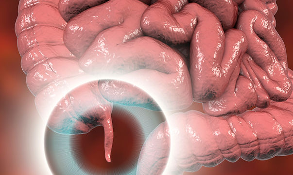
APPENDICITIS
OVERVIEW
Appendicitis is an inflammation of the appendix, a finger-shaped pouch that projects from your colon on the lower right side of your abdomen.
Appendicitis causes pain in your lower right abdomen. However, in most people, pain begins around the navel and then moves. As the inflammation worsens, appendicitis pain typically increases and eventually becomes severe. Although anyone can develop appendicitis, most often it occurs in people between the ages of 10 and 30. Standard treatment is the surgical removal of the appendix.
TYPES OF APPENDICITIS
ACUTE APPENDICITIS
Acute appendicitis is a severe and sudden case of appendicitis. The symptoms tend to develop quickly over the course of one to two days. It requires immediate medical treatment. If left untreated, it can cause your appendix to rupture. This can be a serious and even fatal complication.
Acute appendicitis is more common than chronic appendicitis.
CHRONIC APPENDICITIS
Chronic appendicitis is less common than acute appendicitis. In chronic cases of appendicitis, the symptoms may be relatively mild. They may disappear before reappearing again over a period of weeks, months, or even years.
This type of appendicitis can be challenging to diagnose. Sometimes, it’s not diagnosed until it develops into acute appendicitis.
Chronic appendicitis can be dangerous.
WHAT CAUSES APPENDICITIS?
Appendicitis happens when the appendix gets blocked, often by feces, a foreign body (something inside you that isn’t supposed to be there), or cancer. Once this obstruction occurs, the appendix becomes filled with mucus and swells. This continued production of mucus leads to increased pressures within the lumen and the walls of the appendix. The increased pressure results in blood clotting (thrombosis) and blockage (occlusion) of the small vessels and the slowing/stoppage of the flow of the critical part of the defense system (immune system) - the lymphatic system. At this point, spontaneous recovery rarely occurs. The blockage of blood vessels results in decreased oxygen and nutrient supplies (ischemia) to the tissue /appendix. Ischemia results in tissue and appendix death. The appendix then becomes necrotic. As bacteria begin to leak out through the dying walls, pus forms within and around the appendix (suppuration). The end result is appendiceal rupture (a 'burst appendix') resulting in peritonitis, which may lead to sepsis and eventually death.
SYMPTOMS
Signs and symptoms of appendicitis may include:
-
Sudden pain that begins on the right side of the lower abdomen
-
Sudden pain begins around your navel and often shifts to your lower right abdomen.
-
Pain that worsens if you cough, walk or make other jarring movements.
-
Nausea and vomiting
-
Loss of appetite
-
Low-grade fever may worsen as the illness progresses.
-
Constipation or diarrhea
-
Abdominal bloating
-
Flatulence
The site of your pain may vary, depending on your age and the position of your appendix. When you're pregnant, the pain may seem to come from your upper abdomen because your appendix is higher during pregnancy.

DIAGNOSIS
To help diagnose appendicitis, our board-certified specialists, will likely take a history of your signs and symptoms and examine your abdomen.
Tests and procedures used to diagnose appendicitis include:
-
Physical exam to assess your pain. Our specialists may apply gentle pressure on the painful area. When the pressure is suddenly released, appendicitis pain will often feel worse, signaling that the adjacent peritoneum is inflamed. In addition, our specialists may also look for abdominal rigidity and a tendency for you to stiffen your abdominal muscles in response to pressure over the inflamed appendix (guarding). Finally, our specialists may use a lubricated, gloved finger to examine your lower rectum (digital rectal exam). Women of childbearing age may be given a pelvic exam to check for possible gynecological problems that could be causing the pain.
-
Blood test. This allows our specialists to check for a high white blood cell count, which may indicate an infection.
-
Urine test. Our specialists may want you to have a urinalysis to make sure that a urinary tract infection or a kidney stone isn't causing your pain.
-
Imaging tests. Our specialists may also recommend an abdominal X-ray, an abdominal ultrasound, computerized tomography (CT) scan, or magnetic resonance imaging (MRI)
TREATMENT FOR APPENDICITIS
Appendicitis is almost always treated as an emergency. Surgery to remove the appendix, which is called an appendectomy, is the standard treatment for almost all cases of appendicitis.
Generally, if our specialist suspects that you have appendicitis, they will recommend an immediate appendectomy and quickly remove it to avoid a rupture. If you have an abscess, you may get two procedures: one to drain the abscess of pus and fluid, and a later one to take out the appendix. But some research shows that treating acute appendicitis with antibiotics may help you avoid surgery.
APPENDECTOMY
Before your appendix is taken out, you will take antibiotics to fight infection. You will usually get general anesthesia, meaning you will be asleep for the procedure.
There are two types of appendectomy: open and laparoscopic. The type of surgery performed depends on several factors, including the severity of your appendicitis and your medical history.
OPEN APPENDECTOMY
During an open appendectomy, the surgeon makes one incision in the lower right side of your abdomen. Your appendix is removed, and the wound is closed with stitches. This procedure allows your doctor to clean the abdominal cavity if your appendix has burst.
Our specialists may choose an open appendectomy if your appendix has ruptured, and the infection has spread to other organs. It is also the preferred option for people who have had abdominal surgery in the past.

Laparoscopic Appendectomy
During a laparoscopic appendectomy, the surgeon accesses the appendix through a few small incisions in your abdomen. A small, narrow tube called a cannula will then be inserted. The cannula is used to inflate your abdomen with carbon dioxide gas. This gas allows the surgeon to see your appendix more clearly.
Once the abdomen is inflated, an instrument called a laparoscope will be inserted through the incision. The laparoscope is a long, thin tube with a high-intensity light and a high-resolution camera at the front. The camera will display the images on a screen, allowing the surgeon to see inside your abdomen and guide the instruments. When the appendix is found, it will be tied off with stitches and removed. The small incisions are then cleaned, closed, and dressed.
Laparoscopic surgery is usually the best option for older adults and people who are overweight. It has fewer risks than an open appendectomy procedure and generally has a shorter recovery time.
CLOSTRIDIUM DIFFICILE (aka “C. DIFFICILE” or “C. DIFF”)
C. diff is short for Clostridium difficile, an infectious bacterium that causes symptoms ranging from diarrhea to life-threatening inflammation of the wall of your colon; a condition known as clostridium difficile colitis. It can produce a range of symptoms.
It is estimated to cause half a million illnesses in the United States each year.
About 1 in 6 patients who get C. diff will get it again in the subsequent 2-8 weeks (about 2 months).
Within a month of diagnosis, 1 in 11 people over age 65 died of a healthcare-associated C. diff infection.
WHAT CAUSES C. DIFFICILE?
C. diff exists all around us. It is in the air, water, soil, and the feces of humans and animals.
C. diff bacteria that are outside the body turn into spores that can live on surfaces for weeks or months. These spores are not "active," but they can turn active after you swallow them, and they get into your intestines. Some people have bacteria in their intestines and never have any symptoms, however, for others, the bacteria make toxins that attack the intestines.
A new strain of C. diff bacteria makes larger amounts of toxins. These types of bacteria are hard to treat with medications.
C. diff bacteria spread in health care facilities, like hospitals or nursing homes, where workers are more likely to encounter it and than with patients or residents.
Between 5 to 15 percent of healthy adults — and 84.4 percent of newborns and healthy infants — have C. diff in their intestines, according to the American College of Gastroenterology (ACG). However, other bacteria that live in the intestines usually keep the amount of C. diff under control.
A C. diff infection occurs when there is too much of the bacterium in your intestines.
WHAT SYMPTOMS DOES IT CAUSE?

The main symptom of a C. diff infection is diarrhea. Other symptoms include:
-
abdominal pain or cramps
-
nausea
-
fever
-
loss of appetite
-
dehydration
-
blood in the stool (in severe cases)
Symptoms of a C. diff infection can range from mild to severe. Call our office if you notice you are having diarrhea three or more times a day, or your symptoms are not going away after two or three days.
You should also seek immediate treatment if you have severe abdominal pain or notice blood in your stool.
HOW DOES C. DIFFICILE SPREAD?
The C. diff bacterium comes from feces. You can develop an infection if you touch a contaminated surface and then touch your mouth.
In addition, the spores of C. diff are resistant to many chemicals used for cleaning. As a result, they can stick around for a long time.
WHO IS MOST LIKELY TO GET AN INFECTION?
While anyone can develop a C. diff infection, some people have an increased risk.
Things that can increase your risk include:
-
taking antibiotics, especially a long course of broad-spectrum antibiotics
-
spending a lot of time in hospitals
-
older age
-
having gastrointestinal surgery
-
having a weakened immune system
-
having chronic kidney or liver disease
-
taking proton pump inhibitors (PPIs)
-
prior C. diff infection
HOW IS C. DIFFILE DIAGNOSED?
To diagnose a C. diff infection, our skilled specialists will start by asking some questions about your symptoms and medical history. Next, they may order a stool sample. They can analyze it for toxins or toxin genes of the C. diff bacterium.
If your symptoms are severe, they may also perform a procedure called a sigmoidoscopy.

A sigmoidoscopy, also called a flexible sigmoidoscopy, is a procedure that lets your doctor look inside your sigmoid colon by using a flexible tube with a light on it. It helps your doctor check for:
-
ulcers
-
abnormal cells
-
polyps
-
cancer
Typically, pieces of tissue will be taken as samples to check for any abnormal cell changes.
HOW IS C. DIFFICILE TREATED?
C. diff infections require treatment with antibiotic therapy. If you are already taking an antibiotic for something else, our doctors may have you stop taking it, if possible.
Common antibiotics used to treat C. diff infections are prescribed based on first-line treatments and the severity of the initial infection.
In most cases, you can take the antibiotics by mouth, which is the standard therapy for the three main C.difficile antibiotics. However, some infections may require intravenous (IV) antibiotic therapy.
The Centers for Disease Control and Prevention (CDC) recommends taking an antibiotic course for at least 10 days to treat a C. diff infection.
In the case of someone with recurrent C. diff who has had at least two recurrences after the first episode, a fecal microbiota transplant may be considered as a potential treatment option, after antibiotic therapy.
As you recover, make sure to drink plenty of fluids. Having diarrhea often leads to dehydration, so it is important to replenish the fluids you lose. In more severe cases, you may need intravenous fluid to treat dehydration as well.
In very rare cases, you may need surgery to remove the affected part of your colon.
ARE THERE ANY COMPLICATIONS?
While most C. diff infections don’t cause any long-term problems, more serious ones can lead to complications, such as:
-
Toxic megacolon - swelling and dilation of the large intestine due to inflammation of the intestinal wall and accumulation of excessive amounts of gas.
-
Bowel perforation
-
Kidney injury. In severe cases of C. diff infection, rapid dehydration can lead to acute kidney injury.
IS C. DIFFICILE PREVENTABLE?
Despite its resistance to many cleaning products, there are several things you can do to prevent yourself from developing or spreading a C. diff infection.
Follow these tips to reduce your risk:
-
Wash your hands regularly with soap and warm water. This is especially important after using the bathroom and before eating.

-
Do not take antibiotics unnecessarily. Keep in mind that antibiotics are only effective for bacterial infections and will not treat a viral infection, such as the flu or common cold.
-
Keep surfaces in high-use areas clean. This includes bathrooms and kitchens. Try to periodically clean these areas with products containing bleach. Bleach is effective against the C. diff bacterium.
WHAT’S THE OUTLOOK?
Most C. diff infections respond well to a 10-day course of oral antibiotic treatment.
Once you start taking the antibiotic, you should notice your symptoms start to improve within a day or two. In more severe cases, you may need an IV antibiotic in addition to oral antibiotic therapy.
If you think you have a C. diff infection, contact our office immediately to avoid any complications.
CHOLANGITIS
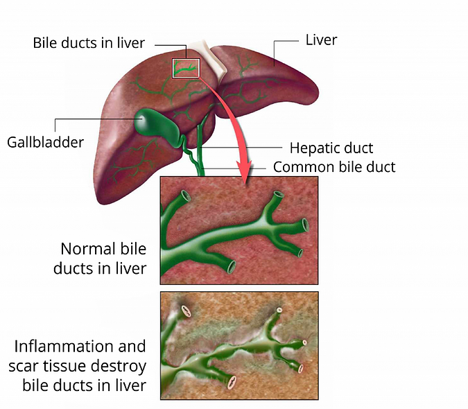
BILE DUCTS AND THE HUMAN LIVER
Your liver is the largest organ inside your body, weighing about 1.4 kg (3 pounds) in an average adult. The liver is in the right upper quadrant of the abdominal cavity, just inferior to the diaphragm in the right superior part of the abdominal cavity and under your right ribs just beneath your right lung – filling much of the right hypochondriac and epigastric regions and extending into the left hypochondriac region. The liver is partially surrounded by the ribs and extends from the level of the fifth intercostal space to the lower margin of the right rib cage, which protects this highly vascular organ from blows that could rupture it. The liver is shaped like a wedge, the wide base of which faces right and the narrow apex of which lies just inferior to the level of the left nipple. The reddish-brown liver is well supplied with blood vessels.
The liver also has two minor lobes, the quadrate lobe, and the caudate lobe. Each lobe is separated into many tiny hepatic lobules, the liver’s functional units. A lobule consists of many hepatic cells radiating outward from a central vein. Blood-filled

channels called hepatic sinusoids separate platelike groups of these cells from each other. Blood from the digestive tract, carried in the hepatic portal vein, brings newly absorbed nutrients into the sinusoids and nourishes the hepatic cells.
OVERVIEW
Cholangitis is a disease in which the bile ducts inside and outside the liver become inflamed and scarred and are eventually narrowed or blocked. Bile ducts carry bile (a fluid that helps to digest fats) from the liver, where bile is produced, to the gallbladder, where it is stored, and to the small intestine, where it aids in digestion.
Cholangitis occurs because of inflammation in the bile ducts (cholangitis) that leads to scarring (sclerosis) and narrowing of the ducts. As a result, bile cannot be released to the gallbladder and small intestine, and it builds up in the liver and causes liver damage. This damage can lead to cirrhosis and eventually liver failure. Medical experts believe that cholangitis is an autoimmune disease, which means that your body’s immune system is overactive and attacks normal, healthy bile duct cells. Cholangitis is strongly associated with inflammatory bowel disease (IBD), particularly ulcerative colitis. Cholangitis is a premalignant condition, associated with higher rates of hepatobiliary and colorectal cancer in patients with ulcerative colitis.
Cholangitis can also be broken down more specifically and known as the following:
-
primary biliary cholangitis (PBC)
-
primary sclerosing cholangitis (PSC)
-
secondary cholangitis
-
immune cholangitis
The bile ducts carry bile from the liver and gallbladder to the small intestine. Bile is a green to yellow-brown fluid that helps your body digest and absorbs fats. It also helps to clear waste from the liver.
When the bile ducts get inflamed or blocked, bile can back up into the liver. This can lead to liver damage and other problems. Some types of cholangitis are mild. Other kinds can be serious and life-threatening.
There are two main types of cholangitis:
-
Chronic cholangitis happens slowly over time. It can cause symptoms over 5 to 20 years.
-
Acute cholangitis happens suddenly. It can cause symptoms over a short time period.
SYMPTOMS OF CHOLANGITIS
Symptoms depend on what kind of cholangitis you have and for how long. Every person with cholangitis may have slightly different signs and symptoms. More than 50 percent of people diagnosed with chronic cholangitis don’t have any symptoms.
Some early symptoms of chronic cholangitis may include:
-
tiredness and fatigue
-
itchy skin
-
dry eyes
-
dry mouth
If you have chronic cholangitis for a long time, you may have:
-
pain in the upper right side
-
night sweats
-
swollen feet and ankles
-
darkening of the skin (hyperpigmentation)
-
muscle pain
-
bone or joint pain
-
bloating (fluid in the stomach area)
-
fat deposits (xanthomas) in the skin around the eyes and eyelids
-
fat deposits in the elbows, knees, palms, and soles of the feet
-
diarrhea or greasy bowel movements
-
clay-colored bowel movements
-
weight loss
-
mood changes and memory problems

If you have acute cholangitis, you may also have other symptoms. These include sudden symptoms like:
-
high fever for more than 24 hours
-
chills
-
nausea
-
vomiting
-
back pain
-
pain below the shoulder blades
-
dull pain or cramps in the upper right side
-
sharp or dull pain in the middle of the stomach
-
low blood pressure
-
confusion
-
yellowing of the skin and eyes (jaundice)
Your doctor may find signs of cholangitis in other parts of the body. These include:
-
swollen or enlarged liver
-
swollen or enlarged spleen
-
high cholesterol
-
underactive thyroid gland (hypothyroidism)
-
weak and brittle bones (osteoporosis)
TREATING CHOLANGITIS
Treatment for chronic and acute cholangitis may be different. This is because the causes of cholangitis vary. Treatment also depends on how early you are diagnosed with cholangitis. Both kinds can lead to serious complications if they are not treated.
Early treatment is especially important for acute cholangitis. Our board-certified specialists' may recommend antibiotics for up to 10 days
They may also recommend procedures in the hospital, such as:
-
intravenous fluids
-
bile duct drainage
Unlike acute cholangitis, no medications are available to treat chronic cholangitis, however, there are is a drug that may help protect the liver. It works by improving bile flow. It does not treat cholangitis itself.
Treatment and care for chronic cholangitis include:
-
managing symptoms
-
monitoring liver function
-
procedures to open blocked bile ducts
Procedures for both chronic and acute cholangitis are:
-
Endoscopic therapy. Balloon dilation may be used to open up the ducts and increase bile flow. This helps to improve and prevent symptoms. You may need endoscopic therapy several times to treat cholangitis. You may have full or local anesthesia (numbing) before the procedure.
-
Percutaneous therapy. This is similar to endoscopic therapy, but it’s through the skin. You will be under localized anesthesia for the procedure.
-
Surgery. The blocked part of the bile duct may be removed. Or, you may have stents put in to open or drain the bile ducts. You’ll be under full anesthesia (asleep) for surgery.
-
Liver transplant. In serious cases, you may need a liver transplant. The damaged liver will be replaced by a new one. You will need to take antirejection drugs for the rest of your life after the surgery. This helps your body keep the new liver healthy.

You may also need treatment for some serious side effects of cholangitis:
-
Nutrition. Cholangitis can affect digestion and how your body absorbs some vitamins. You may need to take vitamin A, D, E, and K supplements.
-
Weak bones. You may be prescribed medications for osteoporosis. Calcium and vitamin D supplements can help improve bone density and strength.
-
High blood pressure. Monitoring and treatment of high blood pressure in the liver (portal hypertension) may be required.
CAUSES OF CHOLANGITIS
There is a range of causes for cholangitis. Sometimes the cause is not known.
Chronic cholangitis may be an autoimmune disease. This means that your body’s own immune system mistakenly attacks the bile ducts. This causes inflammation.
Over time, inflammation can trigger scars or the growth of hard tissue inside the bile ducts. The scarring makes the ducts hard and narrow. They can also block smaller ducts.
Causes of acute cholangitis are:
-
bacterial infection
-
gallstones
-
blockages
-
tumor
Environmental causes of both types of cholangitis include:
-
infections (bacteria, virus, fungi, or parasites)
-
smoking
-
chemicals
RISK FACTORS
Cholangitis frequently occurs secondary to a gallstone obstructing the common bile duct (CBD).
Three major risk factors exist for cholangitis.
Medical history
If you’ve experienced any of the following, your risk of cholangitis is higher:
-
Gallstones, common bile duct (CBD) stones
-
Recent cholecystectomy (gallbladder removal surgery)
-
endoscopic manipulation or ERCP, cholangiogram
-
History of cholangitis
-
History of HIV or AIDS: AIDS-related cholangitis is characterized by extrahepatic biliary edema, ulceration, and obstruction of uncertain cause
Ethnicity
The prevalence of gallstones is highest in fair-skinned people of Northern European descent as well as in Hispanic populations, Native Americans, and Pima Indians.
Certain Asian populations and inhabitants of countries where intestinal parasites are common are also at increased risk. Asians are more likely to have primary stones due to chronic biliary infections, parasites, bile stasis, and biliary strictures. Recurrent pyogenic cholangitis (Oriental cholangiohepatitis) rarely is observed in the United States.
Additionally, Black individuals with sickle cell disease are at increased risk.
Sex and age
Although gallstones are more common in women than in men, the male-to-female ratio is equal in cholangitis.
Elderly patients are more likely to progress from asymptomatic gallstones to serious complications of gallstones and cholangitis. Elderly patients are more prone to gallstones and CBD stones and, therefore, cholangitis.
The median age at presentation of cholangitis is between 50 and 60 years.
Risk factors that might increase your chance of getting cholangitis:
-
Being female. Chronic cholangitis is more common in women.
-
Age. It usually occurs in adults between the ages of 30 and 60.
-
Genetics. Cholangitis may run in your family.
-
Location. The disease is more common in North America and northern Europe.
DIAGNOSING CHOLANGITIS
Our board-certified specialists can diagnose cholangitis with tests and scans. Several signs may show up in the following blood tests:
-
complete blood count (CBC)
-
liver function tests
-
kidney function tests
-
blood culture
You may also have imaging tests including:
-
Ultrasound (also called sonography). This test creates images of your internal organs on a computer screen using high-frequency sound waves. It is used to see organs in your belly such as the liver, spleen, and gallbladder. It also checks blood flow through different vessels. It can be done outside the body (external). Or it may be done inside the body (internal). If internal, it is called an endoscopic ultrasound (EUS).
-
CT scan. A CT scan may be done with a dye that is swallowed or injected through an IV. This will show the abdomen and pelvis including the bile drainage area. It can help determine why there is a blockage.
-
Magnetic resonance cholangiopancreatography (MRCP). This test is used to look for any problems in your abdomen. It can show if there are gallstones in your bile duct. The test is done from outside your body. It does not involve putting a tube (endoscope) into your body. It uses a magnetic field and radio frequency to make detailed pictures.
-
ERCP (endoscopic retrograde cholangiopancreatography). This is used to find and treat problems in your liver, gallbladder, bile ducts, and pancreas. It uses X-ray and a long flexible tube with a light and camera at one end (an endoscope). The tube is put into your mouth and throat. It goes down your food pipe (esophagus), through your stomach, and into the first part of your small intestine (the duodenum). A dye is put into your bile ducts through the tube. The dye lets the bile ducts be seen clearly on X-rays. If required, this procedure can also help open up your bile ducts.
-
Percutaneous transhepatic cholangiography (PTC). A needle is put through your skin and into your liver. Dye is put into your bile duct so that it can be seen clearly on X-rays. This procedure can also be used to open up the bile ducts if your physicians are unable to do it internally with an ERCP.
You might need other tests such as urine, bile, or stool samples.
COMPLICATIONS OF CHOLANGITIS
Cholangitis can lead to serious health problems if it isn’t treated. Complications include:
-
Liver problems. Cholangitis can cause liver scarring (cirrhosis). This can slow liver function or lead to liver failure. It also increases the risk of liver cancer. It can cause liver swelling and high blood pressure.
-
Gallstones. Blocked bile can harden into stones. This may cause pain and infections.
-
Enlarged spleen. If the liver isn’t working properly and can’t filter out wastes and toxins, old blood cells can collect in the spleen, causing it to swell.
-
Enlarged veins. High blood pressure in the liver may put too much pressure on veins in the stomach. This can lead to swollen and broken veins. It may also cause bleeding.
-
Blood infection. Acute cholangitis can lead to sepsis (a blood infection). This can damage several parts of the body and may be life-threatening if not treated.
Chronic cholangitis is also linked to other conditions including thyroid problems, scleroderma, and rheumatoid arthritis.
OUTLOOK
Your signs and symptoms will vary from other people with cholangitis. In some cases, the cause may not be known. You can’t always prevent getting cholangitis.
Early treatment can help you have a better outcome. It also helps to prevent symptoms and complications. See your doctor urgently if you have any symptoms, including:
-
fever
-
abdominal pain
-
yellowing of the eyes and skin
-
changes in digestion and bowel movements
You may not have any symptoms at all. Regular checkups can help you learn about your liver health with a simple blood test.
Some types of cholangitis may be easier to clear up with treatment. Take all medications as prescribed and see your doctor for all follow-up appointments.
You can prevent complications with daily lifestyle changes like quitting smoking. A healthy, balanced diet with plenty of fiber may ease cholangitis symptoms and prevent complications. Talk to your doctor or nutritionist about the best diet plan for you.
CHOLECYTITIS
GALL BLADDER
The gallbladder is a pear-shaped, hollow structure located under the liver and on the right side of the abdomen. It is located in front of the duodenum (the first section of the small intestine) and is approximately an inch wide and 3 inches long,1tapered at one end where it connects to the cystic duct. It has the capacity to store approximately 30 to 50 cubic centimeters (cc) of fluid, called bile.
OVERVIEW
Cholecystitis is a swelling and irritation of the gallbladder. The gallbladders’ function is to store bile until it is needed for digestion. When we eat, the gallbladder contracts, or squeezes, to send bile into your digestive tract.


It releases bile into your small intestine when your body needs it to break down fats. But if the path to your small intestine is blocked, bile gets trapped. That backup can irritate your gallbladder. That is how cholecystitis happens.
If you don’t see a doctor and get treatment, it can lead to dangerous infections or become a long-term condition. The most common solution is surgery to remove your gallbladder.
CHOLECYSTITIS SYMPTOMS
There are two types of cholecystitis:
-
Acute cholecystitis is the sudden inflammation of the gallbladder that causes marked abdominal pain, often with nausea, vomiting, and fever.
-
Chronic cholecystitis is a lower intensity inflammation of the gallbladder that lasts a long time. It may be caused by repeat attacks of acute cholecystitis. Chronic cholecystitis may cause intermittent mild abdominal pain or no symptoms at all. Damage to the walls of the gallbladder leads to a thickened, scarred gallbladder. Ultimately, the gallbladder can shrink and lose its ability to store and release bile.
Gallstones alone can cause episodes of crampy abdominal pain without any infection. This is called biliary colic.
Cholecystitis can mimic other health problems, so you’ll need to see a doctor for a diagnosis.
You might feel a sharp, sudden pain in the upper right side of your belly. You may also feel pain in your back or below your right shoulder blade. Deep breaths may make it worse. Some other symptoms to watch out for include:
-
Bloating
-
Yellow skin or eyes (jaundice)
-
Bowel movements that are loose and light-colored.
Symptoms may get worse after a high-fat meal. If you can’t get comfortable or sit still because your pain is so strong, go to an emergency room.
CHOLECYSTITIS CAUSES
The typical reason bile backs up is that gallstones, lumps of bile turned solid -- block the way to the small intestine. Gallstones are common. About 10% to 20% of Americans have them. About half of people with gallstones will get cholecystitis.

But gallstones aren’t the only problem that can cause this condition. Others include:
-
Gallbladder sludge, a thick liquid, builds up in the organ. This can happen if you’re pregnant or if you’ve lost a lot of weight quickly.
-
Tumors block the bile’s path. A growth in your pancreas or liver can stop it from draining.
-
Your gallbladder doesn’t have a good blood supply. People with diabetes can have this problem.
-
An infection affects your gallbladder. Bacteria can damage the system that drains bile, causing it to back up.
Cholecystitis can come on suddenly. You may hear a doctor or nurse call it an “acute” case. Or it can be a long-term problem. Those cases are called “chronic.”
CHOLECYSTITIS RISK FACTORS
Women are more likely than men to get gallstones. The risk of gallstones also is higher in:
-
Anyone older than age 60
-
Women who are pregnant or have had several pregnancies.
-
Women who take estrogen replacement therapy or birth control pills
-
Obese people
-
People who have lost weight rapidly.
-
People who eat a high-fat diet
You have a higher chance of getting cholecystitis if you:
-
Are a woman older than 50
-
Are a man older than 60
-
Are overweight
-
Have diabetes
-
Are pregnant.
Or if you have:
-
Heart disease
-
End-stage kidney disease
-
Hyperlipidemia (when your blood has too many lipids in it)
-
Lost weight quickly
You also run a bigger chance of getting it if your diet is high in fat and cholesterol or your ancestry is Native American, Hispanic, or Scandinavian.
CHOLECYSTITIS DIAGNOSIS
Our board-certified specialists will examine you, ask a few questions about your symptoms, and probably order some tests. You should be ready to:
-
Detail when your symptoms started. Have you felt this way before?
-
Describe how severe your pain is.
-
Talk about whether anything makes your pain better or worse.
Our specialists can tell from blood tests whether you have an infection and whether your liver is working the way it should. They may also require you to have imaging tests. These may include:
-
X-ray of your belly, which will show your internal organs, bones, and tissues.
-
Ultrasound, which will show your gallbladder and liver and let doctors check blood flow.
-
CT scan, which gives doctors a more detailed look at organs, muscles, and bones
-
HIDA scan, which checks how your gallbladder moves and shows if bile is blocked. You get a shot of a chemical, and then a scanner traces it as it moves through your body.
-
PTC, which uses a dye injected into your liver to show how bile is moving through your body.
-
ERCP, which uses a long, flexible tube threaded down your throat, through your stomach, and into your small intestine. It has a light and a camera at the end. This test also uses a dye to check how bile is flowing through your system.
CHOLECYSTITIS TREATMENT
Treatment for cholecystitis usually involves a hospital stay to control the inflammation in your gallbladder. Sometimes, surgery is needed.
At the hospital, our highly skilled specialists will work to control your signs and symptoms. Treatments may include:
-
Fasting. You may not be allowed to eat or drink at first in order to take the stress off your inflamed gallbladder.
-
Fluids through a vein in your arm. This treatment helps prevent dehydration.
-
Antibiotics to fight infection. If your gallbladder is infected, your doctor likely will recommend antibiotics.
-
Pain medications. These can help control pain until the inflammation in your gallbladder is relieved.
-
Procedure to remove stones. Your doctor may perform a procedure called endoscopic retrograde cholangiopancreatography (ERCP) to remove any stones blocking the bile ducts or cystic ducts.
Your symptoms are likely to decrease in two or three days. However, gallbladder inflammation often returns. Most people with the condition eventually need surgery to remove the gallbladder.
Gallbladder removal surgery is called a cholecystectomy. Usually, this is a minimally invasive procedure, involving a few tiny incisions in your abdomen (laparoscopic cholecystectomy). An open procedure, in which a long incision is made in your abdomen, is rarely required.

The timing of surgery depends on the severity of your symptoms and your overall risk of problems during and after surgery. If you're at low surgical risk, surgery may be performed within 48 hours or during your hospital stay.
Once your gallbladder is removed, bile flows directly from your liver into your small intestine, rather than being stored in your gallbladder. You don't need your gallbladder to live normally.
CHOLECYSTITIS COMPLICATIONS
If you don’t get treatment, your gallbladder can become infected, and some of the tissue may die. Infection can also spread to other parts of your body, including your pancreas (pancreatitis) and the lining of your belly (peritonitis).
If the tubes that carry bile are damaged too much, cholecystitis can harm your liver, too. You could have repeated bouts of painful symptoms. Eventually, your gallbladder will shrink and not work as well. The condition would become a long-term, or chronic, problem.
CHOLECYSTITIS PREVENTION
You can take steps to lower your chances of getting gallstones and cholecystitis. They include:
-
Lower your cholesterol.
-
Exercise regularly.
-
Eat a diet rich in fruits, vegetables, and healthy fats. Eggs, soybeans, and peanuts are great choices.
Obesity is a major risk factor for getting gallstones. Losing weight can reduce your chances but be sure you do it in a healthy way. If you’re planning a rapid weight loss program, such as weight loss surgery, your doctor or nurse should monitor you. They may recommend bile acid pills to prevent gallstones as you lose weight.
DIVERTICULITIS
OVERVIEW
Although it was rare before the 20th century, diverticular disease is now one of the most common health problems in the Western world. It’s a group of conditions that can affect your digestive tract. The most serious type of diverticular disease is diverticulitis.
Diverticular disease develops when pouches form along your digestive tract, typically in your colon (large intestine).
These pouches are known as diverticula. They form when weak spots in your intestinal wall balloon outward.
Diverticulitis happens when diverticula become inflamed and, in some cases, infected. This can occur when feces or partially digested food blocks the opening of the diverticula.
It can cause uncomfortable symptoms and, in some cases, serious complications. If left untreated, these complications can cause long-term health problems.
SYMPTOMS OF DIVERTICULITIS
Diverticulitis can cause symptoms ranging from mild to severe. These symptoms can appear suddenly, or they can develop gradually over several days.
Potential symptoms of diverticular disease include:
-
pain in your abdomen
-
bloating
-
diarrhea
-
constipation
If you develop diverticulitis, you might experience:
-
constant or severe pain in your abdomen
-
nausea and vomiting
-
fever and chills
-
blood in your stool
-
bleeding from your rectum
Abdominal pain is the most common symptom of diverticulitis. It will most likely occur in the lower left side of your abdomen. But it can also develop in the right side of your abdomen. If you develop any of the above symptoms, such as vomiting or blood in your stool, it may be a sign of a serious complication from diverticulitis or

another condition. Call your doctor right away.
WHAT CAUSES DIVERTICULITIS
There’s no single known cause of diverticular disease. Instead, experts believe that multiple genetic and environmental factors likely contribute to its development.
TREATMENT
Treatment depends on the severity of your signs and symptoms.
Uncomplicated diverticulitis
If your symptoms are mild, you may be treated at home. Your doctor is likely to recommend:
-
Antibiotics to treat infection, although new guidelines state that in very mild cases, they may not be needed.
-
A liquid diet for a few days while your bowel heals. Once your symptoms improve, you can gradually add solid food to your diet.
This treatment is successful in most people with uncomplicated diverticulitis.
Complicated diverticulitis
If you have a severe attack or have other health problems, you'll likely need to be hospitalized. Treatment generally involves:
-
Intravenous antibiotics
-
Insertion of a tube to drain an abdominal abscess, if one has formed
Surgery
You'll likely need surgery to treat diverticulitis if:
-
You have a complication, such as a bowel abscess, fistula or obstruction, or a puncture (perforation) in the bowel wall
-
You have had multiple episodes of uncomplicated diverticulitis
-
You have a weakened immune system
There are two main types of surgery:
-
Primary bowel resection. The surgeon removes diseased segments of your intestine and then reconnects the healthy segments (anastomosis). This allows you to have normal bowel movements. Depending on the amount of inflammation, you may have open surgery or a minimally invasive (laparoscopic) procedure.
-
Bowel resection with colostomy. If you have so much inflammation that it's not possible to rejoin your colon and rectum, the surgeon will perform a colostomy. An opening (stoma) in your abdominal wall is connected to the healthy part of your colon. Waste passes through the opening into a bag. Once the inflammation has eased, the colostomy may be reversed and the bowel reconnected.
Follow-up care
A colonoscopy may be recommended six weeks after you recover from diverticulitis, especially if you have not had the test in the previous year. There does not appear to be a direct link between diverticular disease and colon or rectal cancer. But colonoscopy — which is risky during a diverticulitis attack — can exclude colon cancer as a cause of your symptoms.

After successful treatment, our specialists may recommend surgery to prevent future episodes of diverticulitis. The decision on surgery is an individual one and is often based on the frequency of attacks and whether complications have occurred.
COMPLICATIONS
While they are not common, there are several different complications that may occur along with diverticulitis.
Abscess
An abscess is a bacterial infection that causes a pocket of blood and pus to form. Abscesses associated with diverticulitis may cause fever and abdominal pain. They are treated with antibiotics and/or drainage.
Fistula
A fistula is a tunnel that forms in the body and connects either two organs or an organ and the skin.
Symptoms of a fistula (which depends on location) can include a break in the skin, swelling, pain, passing air while urinating, passing stool through the vagina, a visible skin break, or drainage from the area.
A fistula may be treated with surgery or with the use of a seton, which is a thread that is gradually tightened until the fistula is closed.
Bowel Obstruction
A bowel obstruction is a blockage in the intestine which prevents the passage of stool. When diverticulitis leads to a bowel obstruction the symptoms can include abdominal pain, distention, and bloating; constipation or diarrhea; thin stools; and nausea and vomiting.
An obstruction might be treated in the hospital through the use of a nasogastric (NG) tube or in some cases may require surgery.
Perforation
A perforation is a hole in the colon. It is a serious condition that requires treatment immediately in order to prevent complications such as peritonitis, which is a potentially fatal infection.
The symptoms of perforation can include severe abdominal pain, fever, chills, bleeding from the rectum, and nausea and vomiting.
OUTLOOK/ PROGNOSIS
What should I expect if I have been diagnosed with diverticular disease?
If you’ve been told you have diverticulosis, this is usually not cause for concern. This condition is very common and increases with age. It is present in about 50% of people over age 60 and in almost everyone over age 80. You likely won’t even have symptoms if you have diverticulosis. If you have a mild case of diverticulosis, it may go away on its own without treatment.
Up to 30% of people with diverticulosis do develop diverticulitis. Between 5% and 15% will develop rectal bleeding.
Most people who have diverticulitis will recover with about a seven to 10-day course of antibiotics and rest. A severe complication of diverticulitis occurs in about the following percent of people: perforation of the colon (1% to 2% of patients), obstruction (rare), fistula (14%) or abscess (30%).
The best self-treatment is to eat a high-fiber diet (one filled with fruits and vegetables, cereals and whole grains, nuts, beans and legumes. Also, drink more fluids (half your body weight in ounces each day) and exercise (helps speed waste through your colon).
What are the dangers of diverticulitis? Is diverticulitis a life-threatening condition?
Diverticulitis can be a serious, and even a potentially life-threatening complication. Health problems that can arise from diverticulitis include:
-
Rectal bleeding.
-
Abscesses and fistulas.
-
Obstructions and strictures.
-
Perforation, leading to peritonitis.
If I’ve had one bout of diverticulitis, how likely am I to have a repeat bout?
If you’ve had a previous episode of diverticulitis, you have up to about a 20% chance of having a repeat episode. However, fewer than 6% of patients will develop complicated diseases or need emergency surgery.
Can diverticulitis be cured?
Diverticulitis can be treated and be healed with antibiotics. Surgery may be needed if you develop complications or if other treatment methods fail and your diverticulitis is severe. However, diverticulitis is generally considered to be a lifelong condition.
Can I still get diverticulitis if I’ve had the affected part of my colon removed?
If the affected area of your colon is removed, another surgery is usually not needed. The most common location for diverticulitis is the sigmoid colon, which is S-shaped near the end portion of your colon. Although this is the most common location, it’s possible for diverticula to form in other areas of your colon. Because each person is different, be sure to ask your healthcare provider, surgeon, or colon specialist about your risk for return appearance of diverticulitis.
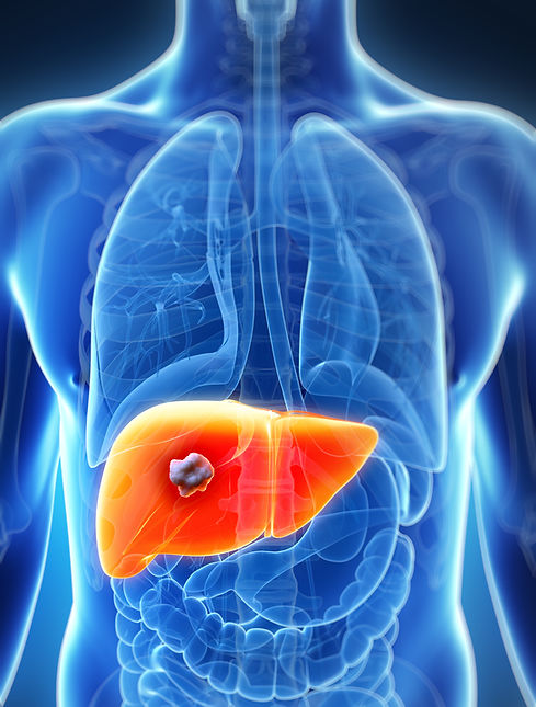
LIVER ABSCESS
The liver, the largest gland in the body, a spongy mass of wedge-shaped lobes that has many metabolic and secretory functions. The liver secretes bile, a digestive fluid; metabolizes proteins, carbohydrates, and fats; stores glycogen, vitamins, and other substances; synthesizes blood-clotting factors; removes wastes and toxic matter from the blood; regulates blood volume; and destroys old red blood cells.
A liver abscess, also known as a hepatic abscess, is an accumulation of pus within the liver as a result of an infection. Overall liver abscesses are fairly rare and more frequently seen in developing nations. Most liver abscesses are caused by bacteria and amebic parasites (protozoa). However, other protozoa, fungi, and helminths (parasitic worms) may also be responsible for hepatic abscesses.
A liver abscess may occur as a single abscess (solitary) or multiple lesions varying in size from a few millimeters to several centimeters in a larger abscess. The term liver or hepatic abscess is also used to refer to a biliary abscess (gallbladder) which is usually multiple. A liver abscess is one type of intra-abdominal abscess and may be associated with other abscesses in the abdomen.
TYPES AND CAUSES OF HEPATIC ABSCESSES
There are several types of liver abscesses based on the causative organism:
Pyogenic abscess – bacteria, one or more species
Amebic abscess – amoebas which are single-celled parasites (protozoa), most common is Entamoeba histolytica.
Fungal abscess – due to fungi, particularly yeasts like the Candida species
Parasitic abscess – rare, associated with helminths.
A pyogenic abscess accounts for the majority of the liver abscesses and is more frequently associated with the following bacteria:
-
E. coli
-
K.pneumoniae
-
S.aureus
-
Streptococci
SIGNS AND SYMPTOMS OF A LIVER ABSCESS
In the past, direct invasion often associated with complications of appendicitis and other intra-abdominal infections were the leading causes of liver abscesses. However better treatment and management these days has reduced the incidence and subsequently ascending infections associated with cholangitis (bile duct infection) has become the most common cause, particularly in the elderly. The signs and symptoms of these conditions may appear at first and therefore should be considered in the early clinical presentation of a liver abscess.
Signs and symptoms of a liver abscess include:

-
Right upper quadrant abdominal pain but may extend to the left side as well or present with referred pain to the right shoulder.
-
Fever and chills.
-
Tenderness in the liver area with an enlargement of the liver (hepatomegaly) palpable.
-
Anorexia – loss of appetite.
-
Nausea
-
Malaise
-
Cough and/or hiccups due to irritation of the diaphragm
-
Jaundice sometimes
Some patients may present with no abdominal pain.
Although rare, a subdiaphragmatic liver abscess may spread into the thoracic cavity to cause empyema (pus around the lungs) or a lung abscess. The clinical features of these conditions may therefore also be present and need to be investigated. Sepsis or peritonitis are other severe complications that need to be considered.
DIAGNOSING A LIVER ABSCESS
The diagnosis of a liver abscess depends on the medical history (history of intra-abdominal infections or signs/symptoms indicating so) and the current clinical presentation. Some patients may only report a fever of unknown origin as the diagnosis could be missed on prior clinical assessment.
Laboratory studies may include a CBC (complete blood count), liver function test (LFT), blood culture, and culture of abscess fluid. A CT scan and ultrasound are usually sufficient to confirm the diagnosis.
TREATMENT OF A LIVER ABSCESS
Antibiotics are usually sufficient for treating small multiple abscesses. Surgical drainage may be required for larger abscesses, especially the solitary massive abscess. Percutaneous (through the skin) drainage may sometimes be considered if suitable. Certain antibiotics like metronidazole may also be used for an amebic abscess and antifungal drugs may be commenced after drainage of a fungal abscess. Without prompt treatment, there is a risk of the infection spreading and the rupture of an abscess has a poor outlook and is often fatal.

PANCREATITIS
WHAT IS PANCREATITIS?
Pancreatitis is pathologic inflammation of the pancreas. Your pancreas sits behind your stomach, near your small intestine. It releases enzymes that help you digest food and regulates how your body manages glucose.
Pancreatitis can come and go quickly, or it can be a chronic problem. Treatment will depend on whether your pancreatitis is acute or chronic.
PANCREATITIS SYMPTOMS
Most people who have acute or chronic pancreatitis experience middle-left upper abdominal pain as their primary symptom. Some people who have chronic pancreatitis may show inflammation on diagnostic imaging scans but otherwise may show no symptoms.
Other symptoms of pancreatitis may include:

-
pain that wraps around the upper body and involves the back in a band-like pattern.
-
Indigestion
-
Nausea or vomiting
-
Abdominal tenderness
-
Unintentional weight loss
-
Bloating with a distended (swollen) abdomen
-
Hiccups
-
Fever
People who have chronic pancreatitis may also experience steatorrhea; fatty stools that give off a foul odor. Steatorrhea can be a sign of malabsorption; you are not getting all your essential nutrients because your pancreas does not secrete enough digestive enzymes to break down your food.
TYPES OF PANCREATITIS
Pancreatitis is generally acute or chronic. Necrotizing pancreatitis can result from extreme cases of acute pancreatitis. Treatment for each case of pancreatitis depends on the severity of symptoms.
Acute pancreatitis
Acute pancreatitis is the main cause of hospital admissions for gastrointestinal issues. According to the National Institute of Diabetes and Digestive and Kidney Diseases (NIDDK), around 275,000 Americans are admitted to the hospital for acute pancreatitis every year.
The onset of acute pancreatitis is often very sudden. The inflammation usually clears up within several days after treatment begins, but some cases could require a hospital stay.
Acute pancreatitis is much more common in adults than in children. Gallstones are the primary cause of acute pancreatitis in adults.
The condition can also develop into chronic pancreatitis, especially if you smoke or regularly drink alcohol.
Chronic pancreatitis
Chronic pancreatitis is an inflammation of the pancreas that comes back consistently or occurs over a long period of time.
People with chronic pancreatitis can have permanent damage to their pancreas and other complications. Scar tissue develops from this continuing inflammation.
Pancreatitis can damage cells that produce insulin; a hormone released by the pancreas that regulates the amount of sugar in your blood. This leads to diabetes in approximately 45 percent of people with chronic pancreatitis.
Long-term alcohol use results in approximately 70 percent of cases of chronic pancreatitis in adults. Autoimmune and genetic diseases, such as cystic fibrosis, can also cause chronic pancreatitis in some people.
Necrotizing pancreatitis
Severe cases of acute pancreatitis can develop into necrotizing pancreatitis, which refers to the death of cells due to disease. This occurs in approximately 10 percent of acute pancreatitis cases, typically when pancreatitis is left untreated.
Inflammation from pancreatitis can cause digestive enzymes to leak into the pancreas. This can result in damage and death of the tissue, leading to necrotizing pancreatitis. Our specialists may order an abdominal ultrasound or CT scan to diagnose the condition.
If you have necrotizing pancreatitis, our specialists may take a sample of the dead tissue to make sure it has not become infected. If you have an infection, you will likely need to take antibiotics and may need to have the dead tissue removed.
The infection of dead tissue increases the risk of death from necrotizing pancreatitis, so it is very important to seek treatment as quickly as possible.
PANCREATITIS CAUSES
Acute and chronic pancreatitis share many of the same causes. These include:
-
Gallstones
-
Drinking a lot of alcohol
-
Some medications
-
Pancreatic cancer
-
Abdominal surgery
-
Infections
-
Cystic fibrosis
-
Injury to your belly
High levels of calcium or triglycerides (a type of fat) in the blood can also lead to chronic pancreatitis.
Gallstones are the most common cause of acute pancreatitis. Gallstones are small, solid masses that form from bile, a fluid that helps with digestion.
A large enough gallstone can get stuck at the junction where the main pancreatic duct and the common bile duct come together. These ducts empty into the duodenum, the first part of the small intestine.
The pancreatic duct carries digestive enzymes from the pancreas. The common bile duct carries bile or other substances from the liver and the gallbladder. A stuck gallstone can cause a backup of these substances, leading to inflammation in both the common bile duct and the pancreas.
PANCREATITIS PAIN
Pain associated with pancreatitis may last from a few minutes to several hours at a time. In severe cases, discomfort from chronic pancreatitis could become constant.
Your pain is likely to increase after you eat or when you are lying down. Try sitting up or leaning forward to make yourself more comfortable.
Activities like yoga, meditation, and acupuncture may help with pain from pancreatitis. You can also try taking pain medication or antioxidant supplements to help relieve pain.
Surgery is currently the last resort for treating pancreatitis, but research indicates that performing surgery earlier during treatment may help with pain relief.
PANCREATITIS COMPLICATIONS
Some people may develop complications. These complications are rare, but they’re more common in people with chronic pancreatitis:
-
Kidney damage
-
Pancreatic cancer
-
Diabetes
-
Malnutrition
-
pancreatic infections
Acute pancreatitis may increase your risk of developing breathing difficulties. It can also cause pseudocysts to form when tissue and other debris collect on your pancreas. These may go away by themselves. If they rupture, it can cause infection and bleeding that can be fatal if untreated.
PANCREATITIS RISK FACTORS
Several factors increase your risk of developing pancreatitis. These include:
-
heavy alcohol use (more than 2 drinks a day)
-
obesity
-
smoking cigarettes
-
genetics
Men are more likely to develop chronic pancreatitis than women.
A combination of risk factors, like smoking and having a family history of pancreatitis, increases your chances of getting pancreatitis. Smoking or drinking alcohol may also increase the risk of acute pancreatitis developing into chronic pancreatitis.
DIAGNOSING PANCREATITIS
The incredible board-certified specialists at Apex Physicians will use a combination of blood tests and imaging studies to make a diagnosis. If you have acute pancreatitis, you will have severe abdominal pain and blood tests may show a significant rise in your level of pancreatic enzymes.
Several types of ultrasound, MRI, and CT scans can reveal the anatomy of your pancreas, signs of inflammation, and information about the biliary and pancreatic ducts. A fecal fat test can also determine if your stools have fat content that is higher than normal.
Pancreatic function test
The pancreatic function test, also called the secretin stimulation test, shows whether your pancreas is responding normally to secretin. Secretin is a hormone that causes your pancreas to release a fluid that helps digest food.

During the test, our specialists will run a tube through your nose or throat and down into your small intestine. They will inject secretin into your vein, then take samples of fluid through the tube.
Our specialists will send the fluid to a lab to help diagnose pancreatitis or other conditions affecting your pancreas.
PANCREATITIS TREATMENT
Treatment for acute or chronic pancreatitis often involves hospitalization. The pancreas is a key contributor to your digestive processes and needs to rest to heal.
For this reason, you may receive specifically tailored fluids and nutrition intravenously (IV) or through a tube that goes from your nose directly into your stomach. This is called a nasogastric feeding tube.
Medication may help control the pain. You may also receive artificial digestive enzymes for chronic pancreatitis if your pancreas is not producing enough of them on its own.
Restarting an oral diet depends on your condition. Some people feel better after a couple of days. Other people need a week or two to heal sufficiently.
Surgery
You may need surgery if other treatments are not working. If you are diagnosed with gallstones, surgery to remove the gallbladder may help. Surgery can also remove diseased parts of your pancreas.
PANCREATITIS PREVENTION
Depending on the cause, you may not be able to prevent pancreatitis. Still, there are several things you can do to reduce your risk:
-
Limit your alcoholic drinks.
-
Stop smoking.
-
Maintain a healthy weight.
-
Eat a balanced diet.
Eating high-fiber foods and avoiding sugar may help you prevent gallstones, which are the main cause of acute pancreatitis.
OUTLOOK
You can control pancreatitis with a healthy lifestyle and medical treatment when necessary. It is particularly important to avoid smoking and drinking a lot of alcohol to reduce your risk of pancreatitis and to help you recover.
If any of your symptoms reappear, please call our office immediately.
PERITONITIS & INTRAPERITONEAL ABSCESSES
OVERVIEW
The peritoneum, large membrane in the abdominal cavity that connects and supports internal organs. It is composed of many folds that pass between or around the various organs. Two folds are of primary importance: the omentum, which hangs in front of the stomach and intestine; and the mesentery, which attaches the small intestine and much of the large intestine to the posterior abdominal cavity.

Peritonitis is inflammation of the peritoneum, the thin layer of tissue covering the inside of your abdomen and most of its organs. The inflammation is usually the result of a fungal or bacterial infection.
Primary peritonitis is also known as spontaneous bacterial peritonitis. It is thought to be the result of bacterial translocation across an intact gut wall. These infections are commonly monomicrobial, and the infecting organism is primarily determined by patient demographics. For example, healthy young girls are most often infected by streptococcal organisms, cirrhotics by gram-negative or enterococcal organisms, and peritoneal dialysis patients by Staphylococcus aureus. Diagnosis requires peritoneal fluid aspiration. Characteristics of infection include white blood cell count (WBC) > 500 cells/mm3, high lactate, and low glucose levels. Positive peritoneal fluid cultures are definitive, and the resolution of infection is marked by a peritoneal fluid with < 250 WBC/mm3.
Secondary peritonitis is caused by microbial contamination through a perforation, laceration, or necrotic segment of the GI tract. Definitive diagnosis is based on clinical examination and history, and specific diagnoses can be confirmed by radiographic imaging. If a patient is stable enough for transport, computed tomography (CT) scan with intravenous and oral contrast is the standard method of evaluating most intra-abdominal pathologies, such as appendicitis, diverticulitis, and colitis. Suspected biliary pathology is the exception, and ultrasound is the preferred initial imaging modality for this spectrum of disease including acute cholecystitis, emphysematous cholecystitis, and cholangitis. Infections associated with secondary peritonitis are commonly polymicrobial and the infecting organisms are those most associated with the source of contamination.
Tertiary peritonitis represents an infection that is persistent or recurrent at least 48 hours after appropriate management of primary or secondary peritonitis. It is more common among critically ill or immunocompromised patients. Because of the poor host defenses, it is also often associated with less virulent organisms, such as Enterococcus, Candida, Staphylococcus epidermidis, and Enterobacter.
Intra-abdominal sepsis is an IAI that results in severe sepsis or septic shock.
Intraperitoneal abscess is an intra-abdominal collection of pus or infected material and is usually due to a localized infection inside the peritoneal cavity. It can involve any intra-abdominal organ or can be located freely within the abdominal or pelvic cavities, including in between bowel loops. IAA is almost always secondary to a preexisting disease process, or concomitant intra-abdominal process. It can be caused by one or multiple bacterial, fungal, or parasitic infectious agents.
CAUSES
Infectious agents including, but not limited to, bacteria and fungi cause peritonitis. Sometimes the infection begins in the peritoneum. More often, the infection spreads from another area of the body. Some of the most common reasons infection could spread to the peritoneum include:
-
Burst appendix.
-
Stomach ulcer.
-
Inflammation of the pancreas (pancreatitis).
-
Severe abdominal injury such as a knife wound.
-
Infection after abdominal surgery.
-
Digestive conditions such as diverticulitis or Crohn's disease.
-
Infection can also translocate from the gut in certain conditions such as liver failure.
Intraperitoneal abscesses sometimes happen because of another condition such as appendicitis or diverticulitis. Many cases, however, happen after surgery.
Abdominal abscesses can be caused by a bacterial infection. The most common bacteria to cause them are found in the stomach and intestines. One of these is Escherichia coli or E. coli. If left untreated, the bacteria will multiply and cause inflammation and kill healthy tissue.
SYMPTOMS
Different people experience different peritonitis symptoms. The most common symptoms are:
-
Severe pain in the abdomen that gets worse when you move.
-
Nausea and vomiting.
-
Fever.
-
Abdomen that is swollen or tender to the touch.
-
Unexplained weight loss.
If you have recently had surgery or trauma to an abdominal organ and have other risk factors, such as diabetes or inflammatory bowel disease, watch signs of an intra-peritoneal abscess.
If you have recently had surgery or trauma to an abdominal organ and have other risk factors, such as diabetes or inflammatory bowel disease, watch signs of an intra-peritoneal abscess.
The peritoneum helps support the organs in the abdominal cavity and also allows nerves, blood vessels, and lymph vessels to pass through to the organs.

Common symptoms are:
-
Fever
-
Belly pain
-
Chest pain or shoulder pain
-
Lack of appetite
-
Nausea and vomiting
-
Change in bowel movements
-
Rectal tenderness or fullness
-
Mass in the belly
-
Malnourishment
DIAGNOSIS
Several other tests can help your doctor diagnose peritonitis:
-
A blood test, called a complete blood count (CBC), can measure your white blood cell count (WBC). A high WBC count usually signals inflammation or infection. A blood culture can help to identify the bacteria causing the infection or inflammation.
-
If you have a buildup of fluid in your abdomen, your doctor can use a needle to remove some and send it to a laboratory for fluid analysis. Culturing the fluid can also help identify bacteria.
-
Imaging tests, such as CT scans and X-rays, can show any perforations or holes in your peritoneum.

CT of the abdominal cavity and pelvis with oral contrast is the leading diagnostic method for suspected intraperitoneal abscesses
TREATMENT
It is important to treat acute infectious peritonitis quickly, so the infection does not spread to other parts of your body and cause more serious
A microbiological culture, or microbial culture, is a method of multiplying microbial organisms by letting them reproduce in predetermined culture medium under controlled laboratory conditions.
health problems. Treatment for peritonitis typically starts with antibiotics to get rid of the infection. Surgery is required to remove infections that seriously damages the peritoneum.
Antibiotics may help treat an infection that could lead to an intra-abdominal abscess. But once the abscess has developed, antibiotics don't work as well. An abscess often will need to be drained of fluid to heal. But often antibiotics are given along with draining the abscess. The type of antibiotic will depend on how severe your abscess is, your age, and any other health problems you may have.
SPLENIC ABSCESS
OVERVIEW OF THE SPLEEN
The spleen is an organ in the upper far left part of the abdomen, to the left of the stomach. The spleen varies in size and shape between people, but it’s commonly fist-shaped, purple, and about 4 inches long. Because the spleen is protected by the rib cage, you can’t easily feel it unless it’s abnormally enlarged.
The spleen plays multiple supporting roles in the body. It acts as a filter for blood as part of the immune system. Old red blood cells are recycled in the spleen, and platelets and white blood cells are stored there. The spleen also helps fight certain kinds of bacteria that cause pneumonia and meningitis.
SYMPTOMS
The history and physical examination are not sufficiently reliable to make the diagnosis of splenic abscess. However, information derived from the history and physical examination can suggest this diagnosis. Clinicians must maintain a high index of suspicion, particularly in higher-risk clinical scenarios and patient groups.
Although the signs and symptoms of splenic abscess have been well described, they are not very specific. Therefore, splenic abscess remains a substantial diagnostic challenge. The classic triad of fever, left-upper-quadrant pain, and splenomegaly is seen in only about one-third of patients.
The symptoms of a splenic abscess can be variable and depend on the location, size, and progression of the process. They can also be acute, subacute, or chronic. Deep-seated, small abscesses can be painless and accompanied by septic symptoms. The following may be noted:
-
Fever (>90%) can be moderate, continuous, intermittent, or even absent
-
Abdominal pain (>60%) typically occurs suddenly, with a punctum maximum in the left hypochondrium (>39%); pain usually signifies perisplenitis
-
Involvement of the diaphragmatic pleura can cause shoulder pain; the associated eponym is the Kehr sign, though there is no clear demonstration that Kehr either described it or suffered from it
-
Pleuritic chest pain around the left lung base (>15%) is aggravated by coughing or forced expiration
-
General malaise and other constitutional and dyspeptic symptoms can be included, all of which can also be seen in a variety of other septic conditions
WHAT CAUSES A SPLENIC ABSCESS?
Almost all splenic abscesses develop as a result of bacteremia (or fungemia) although a small percentage spread to the spleen from a contiguous site. Endocarditis is the most classic underlying condition that results in splenic abscess although urinary tract infection, surgical wound infections, and gastrointestinal infections that result in bacteremia also lead to a splenic abscess. In some cases, an abscess elsewhere in the abdomen may communicate and involve the spleen. It is known as pancreatic abscesses, and diverticulitis may sometimes extend and involve the spleen.
Bacteremia is the most common predisposing risk factor. Classically, endocarditis is associated with splenic abscess but any cause of bacteremia, ranging from typhoid fever to line infection, to urinary tract infection, may result in a splenic abscess.
Abnormalities of the spleen, either primary or secondary, are risk factors. For example, splenic trauma accidental or medically induced, accounts for as many as 30% of splenic abscesses, although in more recent series, trauma is less frequently a predisposing factor.
Felty syndrome and amyloidosis predispose to a splenic abscess. Other factors include hemoglobinopathies, intravenous drug use, and diabetes mellitus.
The frequency of immunosuppression, particularly human immunodeficiency virus (HIV) infection but also cancer chemotherapy and steroid use, as a predisposing factor in a splenic abscess, appears to be increasing and appears to be present in nearly a third of cases.
ETIOLOGY
Splenic abscesses have diverse etiologies. The most common is hematogenous spread originating from an infective focus (most commonly involving aerobes) elsewhere in the body.
Infective endocarditis, a condition associated with systemic embolization in 22-50% of cases, has a 10-20% incidence of associated splenic abscess. Other infective sources include typhoid, paratyphoid, malaria, urinary tract infection (UTI), pneumonias, osteomyelitis, otitis, mastoiditis, and pelvic infections.
Organisms associated with splenic abscess include the following:
-
Aerobes (in most published cases) - Gram-positive cocci (Streptococcus, Staphylococcus, and Enterococcus [predominant in most reports]); gram-negative bacilli (Escherichia coli, Klebsiella pneumoniae, Proteus, Pseudomonas, and Salmonella [occasionally predominant])
-
Anaerobes - Peptostreptococcus, Bacteroides, Fusobacterium, Clostridium, and Propionibacterium acnes
-
Polymicrobial (up to 50% of cases)
-
Fungi - Candida
%20.jpg)
-
Unusual flora - Burkholderia pseudomallei (occasionally reported in melioidosis); actinomycetes and mycobacteria (most typically seen in immunosuppressed patients)
Pancreatic, other retroperitoneal, and subphrenic abscesses, as well as diverticulitis, may contiguously involve the spleen. Splenic trauma is another well-recognized etiologic factor. Splenic infarction resulting from systemic disorders, such as hemoglobinopathies (especially sickle cell disease), leukemia, polycythemia, or vasculitis, can become infected and evolve into splenic abscesses.
EPIDEMIOLOGY
Splenic abscesses are uncommon and have only been reported in small numbers in the medical literature. Splenic abscesses are uncommon, and their incidence in various autopsy series is estimated at ~0.4% (range 0.14-0.7%). The frequency may be rising due to a rise in the use of immunosuppressive agents and prevalence of immunosuppressive states, higher survival of leukemic patients, and high incidence of drug abuse
TREATMENT AND PROGNOSIS
Admission is recommended for all patients with a splenic abscess. High-dose parenteral broad-spectrum antibiotics are of paramount importance, while further diagnostic and therapeutic arrangements are made. The culture results guide the choice of antibiotics.
The gold standard for treating a splenic abscess is splenectomy. There are two different procedures to perform a splenectomy:
-
Laparoscopic splenectomy &,
-
Open Splenectomy Surgery
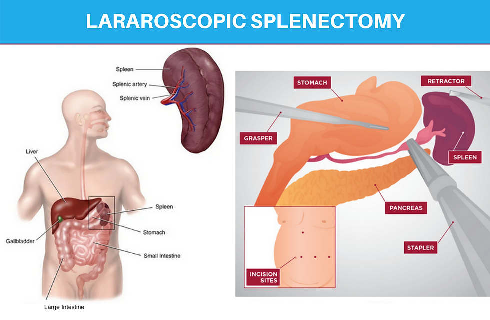
Laparoscopic splenectomy is performed using an instrument known as a laparoscope. It’s a very thin piece of equipment with a micro-mini camera and a light on the end. Under Laparoscopic splenectomy, surgeon 3-4 small incision on the abdomen and inserts the Laparoscope to view inside your abdomen to locate the spleen. The surgeon removes the spleen from surrounding structures and the blood supply of the body. Once the spleen is removed surgeon closes the incision with sutures.
In a rare condition, the surgeon might have to switch from laparoscopic splenectomy to an open procedure. This usually happens when you have excessive bleeding problems during the surgery.
Open splenectomy
In open splenectomy, the surgeon makes a large incision as an entry point to the center or left side of the abdomen underneath the rib cage. After identifying the spleen, the surgeon removes the spleen from the pancreas and blood supply of the body and finally removes the spleen from the body. Once the spleen is removed the surgical incision is closed with stitches or sutures.
Less invasive approaches
Recent studies have shown success using different approaches based on abscess characteristics. Percutaneous aspiration may be a less invasive option in patients at high risk for surgery or a temporary solution used as a bridge to surgery, avoiding the risk of a fulminant and potentially life-threatening infection. A percutaneous aspiration is a successful approach when the abscess collection is unilocular or bilocular, with a complete and thick wall and no internal septations. Aspiration is easier to achieve when the content is liquid enough to be drained. If there are multiple collections or associated coagulopathy, either laparoscopic or open surgical treatment is preferred.
Percutaneous drainage is less likely to be successful in patients with multilocular abscess, ill-defined cavities, necrotic debris, and thick, viscous fluid. Contraindications for percutaneous drainage include the following:
multiple small abscesses, debris-filled cavities, coagulopathy, poorly defined cavities, diffuse ascites, difficult access. Medical treatment alone is not recommended and remains a controversial subject. Mortality rates of more than 50% have been reported in patients only managed with antibiotics. One should consider fungi, actinomycetes, or Mycobacterium as a cause in patients who do not respond. Fungi are known to respond well to antifungal treatment alone. One study also noted that corticosteroid therapy in these patients could be beneficial. Open drainage is sometimes required when percutaneous drainage fails. The routes for open drainage include abdominal intraperitoneal, trans pleural, retroperitoneal.
Splenic abscess is not a frequent clinical problem. However, if the diagnosis is missed, splenic abscess does carry very high mortality reaching more than 70% with appropriate treatment, the mortality can be reduced to less than 1%. Unlike in the past, the prognosis of a splenic abscess today is markedly improved. The availability of percutaneous CT-guided drainage is not only safe and less invasive, but it also avoids the morbidity of open surgery in suitable patients. Furthermore, laparoscopic splenectomy has been a promising alternative to the open method, with faster recovery and short hospital stays. Surgical splenectomy is the treatment of the last choice since most cases can be managed with percutaneous guided drainage and antibiotics.
TYPHLITIS
OVERVIEW
Cecum
Typhlitis refers to inflammation of a part of the large intestine known as the cecum. The cecum or also known as “caecum,” is a small intraperitoneal pouch-like structure located on the right side of the lower abdomen.
It is the starting point of the large intestine and lies between the ileum and the ascending colon, into which the ileum (last part of the small intestine) empties digested content from the small intestine, to be further processed and digested in the large intestine.
Cecum helps in absorbing water from the solid waste produced by the small intestine and also lubricates and further processes the waste with the help of various enzymes to be digested in the large intestine and through the end stage of digestion.
Various functions performed by the cecum are:
Liquid Receptacle: The small intestine absorbs all the nutrients from the food and transfers the solid and liquid waste towards the large intestine. The cecum is the first part of the large intestine that helps in receiving the liquid waste and helps in absorbing liquid from the wastage.
Absorbing Salts: Apart from being a reservoir for liquid waste the cecum also helps in absorbing the salts and electrolytes from the liquid food waste.
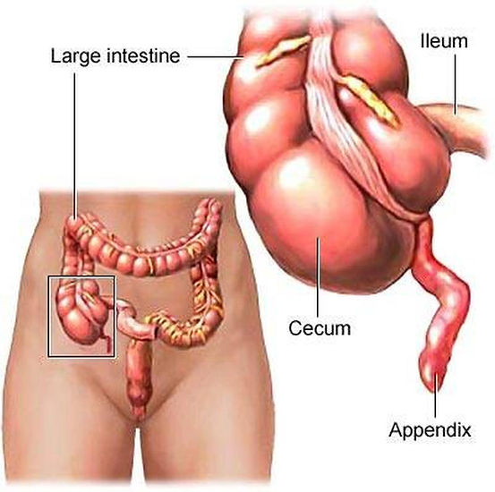
Lubrication: The lining of the cecum has numerous mucous-producing cells that help in lubricating and transferring the solid waste through the large intestine.
Cellulose Breakdown: Cecum also has essential cellulose-digesting enzymes, which help in breaking the cellulose fibers.
Typhlitis is a severe condition that usually affects people with a weakened immune system. They cannot fight off infections like people with healthy immune systems. Typhlitis may also be called neutropenic enterocolitis, necrotizing colitis, ileocecal syndrome, or cecitis.
Typhlitis mostly affects those receiving very intensive chemotherapy drugs to treat cancer. While the exact cause of typhlitis isn’t completely understood, the condition generally occurs when the intestine is damaged, usually as a side effect of the chemotherapy treatment. The intestinal damage along with the person’s weakened immune system makes them more vulnerable to serious infections. These infections can be fatal.
The signs and symptoms of typhlitis are similar to those of a severe intestinal infection.

The symptoms often come on suddenly and include:
-
nausea
-
vomiting
-
chills
-
high fever
-
diarrhea
-
stomach pain or tenderness
-
bloating
People undergoing chemotherapy may also have neutropenia. Neutropenia is a side effect of chemotherapy. It occurs when the immune
system has abnormally low levels of neutrophils, a type of white blood cell important for fighting off infections. Symptoms will often appear around two weeks following a course of chemotherapy.
CAUSES
Researchers believe that typhlitis occurs when the lining of the intestine (mucosa) becomes damaged. This damage is typically caused by a chemotherapy drug. It’s thought that most cases of typhlitis in adults are due to the increasing use of a specific type of cancer treatment known as cytotoxic chemotherapy.
The damaged intestine is then invaded with opportunistic bacteria or fungi. Normally, a person’s immune system would react to this invasion and kill the microorganism. However, people who are immunocompromised won’t be able to fight off the infection.
Typhlitis is most commonly reported in people with the following conditions:
-
leukemia (most common), a cancer of the blood cells
-
lymphoma, a group of cancers that begins in cells of the immune system.
-
multiple myeloma, a type of cancer that affects plasma cells found in the bone marrow.
-
aplastic anemia, a form of anemia where the bone marrow stops making blood cells.
-
myelodysplastic syndromes, a group of disorders that causes low levels of red blood cells, white blood cells, and platelets.
-
HIV or AIDS, a virus that destroys cells of the immune system known as T cells.
-
It’s also reported in people who’ve received a solid organ or bone marrow transplant.
TREATMENT
Typhlitis is a medical emergency and requires treatment right away. Doctors have not yet determined the best way to manage typhlitis.
Currently, treatment involves prompt administration of IV antibiotics, general supportive care (such as intravenous fluids and pain relief), and bowel rest. Bowel rest is when you aren’t allowed to eat or drink anything. Instead, you receive fluids and nutrients through a tube connected to a vein. A suction tube may also be placed through the nose into the stomach to help keep the stomach emptied of digestive juices.
Emergency surgery may be needed to treat complications, such as hemorrhage and a bowel perforation. However, surgery in people with neutropenia can be very risky and may be delayed, if possible, until the neutropenia has improved.
If the typhlitis was caused by a specific type of chemotherapy, later courses of chemotherapy might require a change to a different agent.
COMPLICATIONS
The inflammation can spread to other parts of the intestine. If the blood supply is cut off to the intestine because of the swelling and injury, the tissues may die (necrosis). Other complications include the following:
-
Bowel perforation: when a hole forms all the way through the intestine
-
Peritonitis: Inflammation of the tissue that lines the abdominal cavity.
-
Intestinal bleeding (hemorrhage): bleeding into the intestine.
-
Intestinal obstruction: when the intestine becomes partially or fully blocked.
-
Intra-abdominal abscess: a pocket of inflamed tissue filled with pus caused by an infection that enters the abdomen.
-
Sepsis: a life-threatening infection of the bloodstream.
-
Death
OUTLOOK
The prognosis for typhlitis is generally very poor. One research paper found that the mortality rate could be as high as 50 percent in people with typhlitis. Those who are able to recover faster from a low white blood cell count tend to have better outcomes. Though not common, typhlitis can recur even after treatment.
Early diagnosis and aggressive treatment for typhlitis is needed for a good outcome, but advances in medical technology are expected to improve outcomes in the future.
VIRAL HEPATITIS
OVERVIEW
Hepatitis involves inflammation of the liver cells and damage to the liver. There are different types and causes of hepatitis, but the symptoms can be similar.
The liver is essential for removing toxins from the blood, storing vitamins, and producing hormones. Hepatitis, however, can disrupt these processes.
The most common causes of viral hepatitis are the five unrelated hepatotropic viruses Hepatitis A, B, C, D, and E. The three most common are hepatitis A, B, and C. Infection with any of these three viruses can lead to life-threatening complications.
Each type has different characteristics, and transmission happens in different ways, but the symptoms tend to be similar.
-
Hepatitis A: According to the Centers for Disease Control and Prevention, there were about 2,007 instances of acute hepatitis A infections in the U.S. in 2016. This form of hepatitis does not lead to chronic infection and usually has no complications. The liver usually heals from hepatitis A within several months. However, occasional deaths from hepatitis A have occurred due to liver failure, and some people have required a liver transplant for acute hepatitis A infection. Hepatitis A can be prevented by vaccination.
-
Hepatitis B: Around 22,000 new cases of hepatitis B occurred in 2017, and around 900,000 people are living with the disease in the US. Approximately 95% of adults recover from hepatitis B and do not become chronically infected. However, a few cases cause a life-long, chronic infection. The earlier in life hepatitis B is contracted, the more likely it is to become chronic. People can carry the virus without feeling sick but can

still spread the virus. Hepatitis B can be prevented by getting a vaccine.
-
Hepatitis C: Hepatitis C is one of the most common causes of liver disease in the U.S. and used to be the number one reason for a liver transplant.
About 75% to 85% of patients with hepatitis C develop a chronic liver infection. Roughly 2.4 million people in the U.S. are estimated to have chronic hepatitis C infection. It often does not show any symptoms. No vaccine is yet available to prevent hepatitis C.
-
Hepatitis D: Hepatitis D only happens to people who are infected by the hepatitis B virus. If you are vaccinated against hepatitis B, you will be protected against the hepatitis D virus.
-
Hepatitis E: This type of hepatitis is spread by ingesting contaminated food or water. Hepatitis E is common throughout the world. Even though vaccines exist, they are not available everywhere.
Apex ID specialists are adept at determining and identifying the virus causing hepatitis given that other viruses, such as CMV, EBV, and HSV can also cause hepatitis.
Most people recover from hepatitis, and the disease is often preventable. However, it is still considered a serious health risk because it can:
-
Destroy liver tissue.
-
Spread easily from person to person.
-
Weaken the body's immune system.
-
Cause the liver to fail.
-
Cause liver cancer.
-
Cause death (in rare cases).
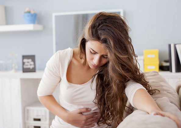
The most common symptoms of hepatitis include:
-
Dark urine.
-
Stomach pain
-
Yellow skin or eye whites called jaundice.
-
Pale or clay-colored stool
-
Low-grade fever
-
Loss of appetite.
-
Fatigue.
-
Feeling sick to your stomach.
-
Aching joints.
WHAT ARE THE SYMPTOMS OF HEPATITIS?
HOW IS HEPATITIS TRANSMITTED?
Hepatitis A can be spread through food or drinking water carrying the virus through bits of fecal matter from an infected person. (This is called the fecal-oral route.) You can also get hepatitis A from sexual contact.
A person can get hepatitis B in many ways, including:
-
Having sex with an infected person.
-
Sharing dirty needles.
-
Being in direct contact with infected blood.
-
Getting needle stick injuries.
-
Being transferred from mother to unborn child.
-
Being in contact with an infected person's body fluids.
An infected mother has a high chance of giving hepatitis B to her child during or after birth. All pregnant women should be tested for hepatitis B. Within 12 hours of birth, infants born to mothers with hepatitis B need to receive treatment with hepatitis B antibody and hepatitis B vaccine. This can prevent the transmission of hepatitis B from the mother to the baby.
A person can get hepatitis C from:
-
Sharing dirty needles.
-
Being in direct contact with infected blood.
-
Getting needle stick injuries.
-
Having sex with an infected person (less common).
Blood products are currently tested for hepatitis B and C, so it is not likely that a person will get hepatitis from receiving them. However, blood transfusions or organ transplants before 1992 might have not been tested for hepatitis (in particular, hepatitis C). If you received a procedure before 1992, you might want to get tested for hepatitis.
Additionally, “baby boomers,” born in the U.S. between 1945 and 1965, are at increased risk of having hepatitis C. If you are part of this group, you should be screened for hepatitis C even in the absence of risk factors listed above. The US Preventive Services Task Force expanded the one-time screening population for hepatitis C to adults from the ages of 18-79, but this recommendation has not been finalized.
You can get hepatitis D from:
-
Being passed from mother to child during childbirth.
-
Having contact with infected body fluids or blood.
You can only get hepatitis D if you have hepatitis B. Hepatitis D is not common in the U.S.
You can get hepatitis E by eating or drinking food or water contaminated with the virus (the fecal-oral route). You can also be infected from under-cooked foods like pork, venison, or shellfish. Hepatitis E is uncommon in the U.S. but can occur after travel to a country where this infection is common. Hepatitis E can be particularly dangerous and even fatal in pregnant women.
RISK FACTORS FOR HEPATITIS INFECTIONS?
You are at a higher risk of getting hepatitis if you:
-
Share needles to take drugs.
-
Practice unprotected oral and/or anal sex.
-
Have many sex partners.
-
Drink significant amounts of alcohol.
-
Have poor nutrition.
-
Work in a hospital or nursing home.
-
Receive long-term kidney dialysis.
-
Travel to areas with poor sanitation.
DIAGNOSING HEPATITIS

The symptoms of the different types of hepatitis are similar, but laboratory tests can identify the specific type a person has.

Our board-certified specialists will perform a physical examination and ask questions to find out about a person’s possible exposure to hepatitis.
They may recommend blood tests or nucleic acid tests. Blood tests can detect antibodies and assess liver function, while nucleic acid tests can — for hepatitis B and C — confirm the speed at which the virus is reproducing in the liver, which will show how active it is.
TREATMENT
There are no treatments to cure hepatitis A, aside from carefully monitoring liver function. If you know you have hepatitis A early enough, you might be able to stop the infection if you get a dose of the hepatitis A vaccine or something called hepatitis A immune globulin.
Hepatitis B, when chronic, can often be treated successfully.
For hepatitis C
These new drugs are sometimes given with older drugs like ribavirin and peginterferon alfa-2a and peginterferon-2b. You might have to take these medicines for some time, even as long as six months.
If you have chronic hepatitis D, our specialists may prescribe drugs with interferons and may also add medicines for hepatitis B. Hepatitis E treatments include peginterferon alfa-2a and ribavirin.
HEPATITIS PREVENTION
There are many ways you can reduce your chances of getting hepatitis:
-
Get the vaccines for hepatitis A and hepatitis B.
-
Use a condom during sex.
-
Don't share needles to take drugs.
-
Practice good personal hygiene such as thorough handwashing with soap and water.
-
Don't use an infected person's personal items.
-
Take precautions when getting any tattoos or body piercings.
-
Take precaution when traveling to areas of the world with poor sanitation. (Make sure to get your vaccines.)
-
Drink bottled water when traveling.

It is very important that you take these preventive measures if you participate in risky behaviors. Take preventive steps, too, if you work in places like nursing homes, dormitories, daycare centers, or restaurants where there you have extended contact with other people and risk of coming into contact with the disease.
OUTLOOK FOR HEPATITIS
Hepatitis A and E usually only cause short-term (acute) infections that your body can overcome. The others (B, C, and D) can also cause acute infections, but might also cause chronic (long-term) infections. The chronic forms are more dangerous. Hepatitis non-E is usually acute but can become chronic.
Most people recover fully from hepatitis even though it might take several months for the liver to heal. To help improve your health and to help speed up your recovery:
-
Avoid alcohol.
-
Practice good nutrition.
-
If you feel sick, rest.
-
Talk to your healthcare provider about your medicines, even over-the-counter drugs or vitamins and supplements, to know which ones you should take and which to avoid until you are recovered.
With hepatitis, your healthcare provider will also be looking for long-term damage to the liver in the forms of cirrhosis or liver failure. You may be asked to take other types of tests, such as liver function tests, imaging tests or possibly a liver biopsy.
If you have questions, new symptoms, or worsening of any existing symptoms, you should call the office of your healthcare provider.
In the U.S., A, B and C are the most common viral forms of hepatitis. It doesn’t matter how you were infected—what matters is taking care of yourself once you have been diagnosed and taking care not to spread the infection to anyone else.